Scientists: Targeting Vaults to Cell Surface Receptors
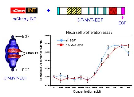
Vaults have been engineered with externally-oriented peptides and protein domains to allow cell type specific targeting via receptor-mediated endocytosis (Kickhoefer et al.,2009). Three different tags have been engineered onto the C-terminus of MVP: an 11 amino acid epitope tag from vesicular stomatitis virus (VSVG), a 33 amino acid IgG-binding peptide (Z domain), and the 55 amino acids encoding epidermal growth factor (EGF). These modified vaults were produced using the baculovirus expression system. The figure below shows a schematic model for the EGF vault which was packaged with the red fluorescent protein, mCherry (produced as a fusion protein with the INT targeting domain). The vaults expressing EGF on their surface (called cp-MVP-EGF, see model) were tested for biological activity using a HeLa cell proliferation assay comparing equimolar amounts of recombinant human EGF (rhEGF) with the recombinant vaults displaying EGF. The results indicated that EGF attached to vaults was as bioactive as the recombinant human EGF.
The externally-oriented vault tags can be used to specifically bind the modified vaults to A431 epithelial cancer cells via the epidermal growth factor receptor (EGFR), either directly (EGF modified vaults) or as mediated by a monoclonal antibody (anti-EGFR) bound to recombinant vaults containing the IgG-binding peptide. The EGF modified vaults have the ability to specifically bind cell surface receptors and trigger receptor activation in a manner similar to recombinant human EGF, initiating a cascade of signaling pathways demonstrated by increased cell proliferation and autophosphorylation of tyrosine 1173 on EGFR (see Figure below). The ability to target vaults to specific cells represents an essential advance towards using recombinant vaults as delivery vehicles.
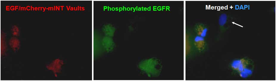
EGF vaults are bioactive. Serum-starved A431 cells were treated with 200 micrograms/mL of purified EGF vaults containing mCherry-INT (red). Following vault binding, the cells were immunostained with the antiphosph-EGFR (Tyr1173) antibody conjugated to Alexa Fluor 488 (green). Merged images show coincident staining as yellow, indicating that only those cells that bound to the EGF/mCherry-INT vaults contain phosphorylated EGFR. The white arrows indicate cells in the field that the vaults did not bind to; therefore, they do not contain phoshorylated EGFR (Kickhoefer et al.,2009).