Big Kids: History

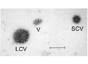
In the mid 1980's Professor Leonard Rome and his colleagues at the UCLA School of Medicine were studying the movement of proteins within cells from their site of production to their final destination (which was the lysosome). These proteins are shuttled in cells in a specialized container called a coated vesicle. Dr. Nancy Kedersha, a researcher working in Dr. Rome's laboratory, was separating coated vesicles into highly-purified fractions. The purification of the vesicles was followed by examining the fractions under the electron microscope using negative staining. One population of coated vesicles was contaminated with an unusual ovoid particle that was highly regular in its form and dimensions. A microscope image from this fraction is shown here.
The figure at left shows a negative stained TEM image from a coated vesicle preparation. Present is a large coated vesicle (LCV), a small coated vesicle (SCV) and an unusual particle which possessed a complex knotted structure (V). The bar represents 100 nmeters.
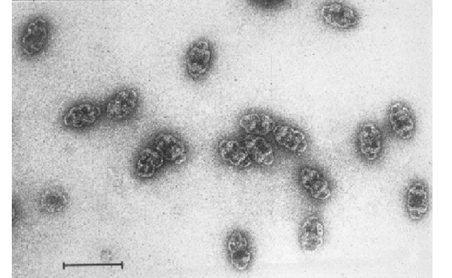
Dr. Kedersha modified the purification procedure and was able to separate the unusual ovoid particles away from the coated vesicles. The purified particles are shown in the photo below.
The figure above shows an image of purified vaults. The bar (lower left) is 100 nmeters (a nmeter is one millionth of a meter).
The purified ovoids were examined using standard biochemical methods which revealed that the particle consisted largely of a single protein with a molecular weight of 104,000 daltons (this size is about average for a protein, the common blood protein hemoglobin is composed of 60,000 dalton subunits). Further analysis established that the particles were unrelated to coated vesicles and thus novel. "Vaults" was chosen as the name for the particle, a term selected to describe the structural features which consists of multiple arches reminiscent of those that form vaulted ceilings of cathedrals.
