Big Kids: Delivering Vaults to the Cytoplasm
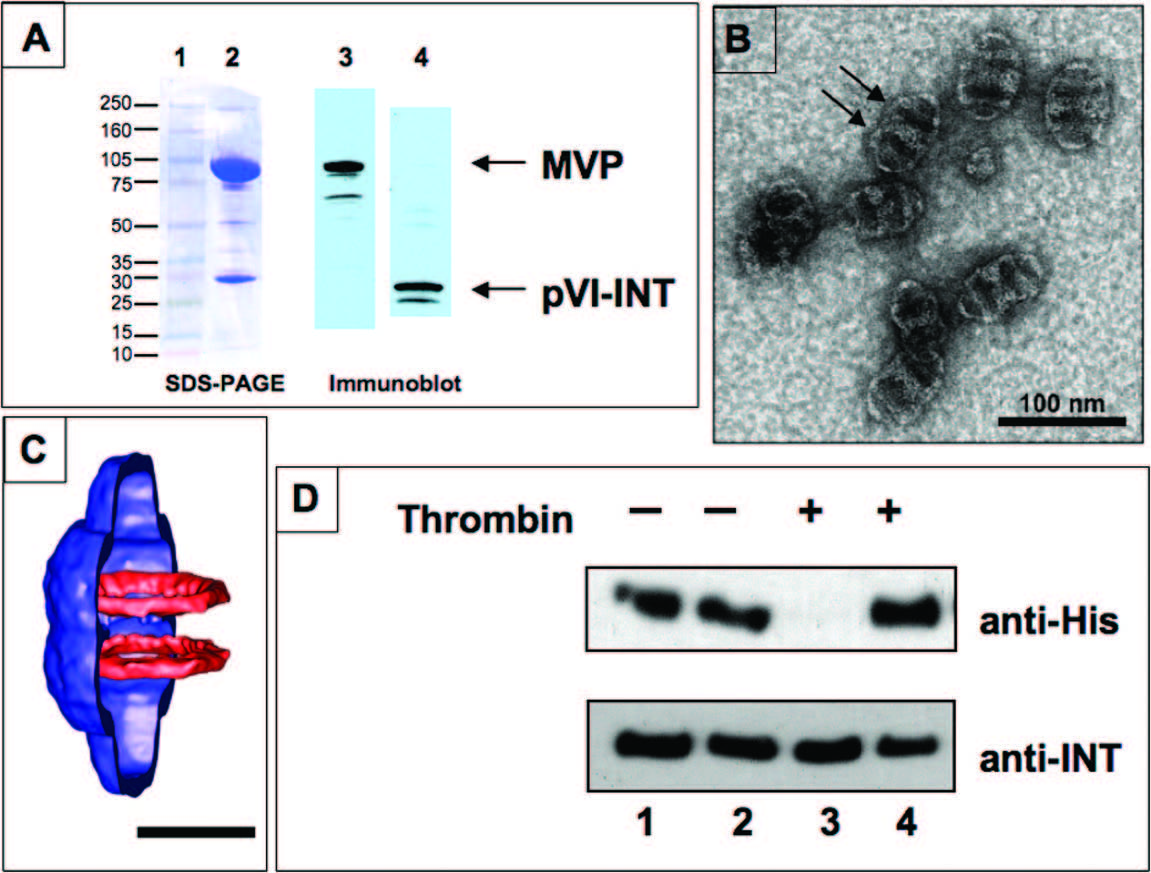
Vaults delivered to cells by binding to cell surfaces will enter the cells by a process called endocytosis. A portion of the cell surface invaginates and pinches off inside the cell to form a structure called an endosome. In order to design the vault as a flexible therapeutic delivery vehicle, it will be important to use the particle to deliver contents to the cyplasmic compartment, therefore getting the vault out of the endosome is an important goal. In collaboration with Glen Nemerow's laboratory at Scripps Research Institute, the Rome Laboratory has analyzed the ability of vaults to directly escape the endosome using a protein derived from a virus (adenovirus protein VI (pVI)) (Lai et al. ACS Nano 3, 691-699 (2009)). This protein has the ability to break open the endosome membrane and allow the contents to get into the cell cytoplasm. To explore the feasibility of enabling vault entry into the cytoplasm of target cells, the membrane breaking domain of adenovirus pVI was incorporated into the interior of recombinant vault particles via fusion to the INT domain to make pVI-INT. As see in the figure below in A, recombinant vaults which were packaged with a pVI protein composed of amino acids 34-114 of pVI fused to the VPARP INT domain were purified, and fractionated on a gel and stained. The purified vaults are shown stained with a blue protein stain in lane 2. Detection of purified vaults was achieved using either anti-MVP monoclonal antibodies (lane 3) or with rabbit polyclonal antisera directed against adenovirus pVI (lane 4). These results confirm the pVI protein was fused with the INT domain and this fusion protein was packaged into the recombinant vaults. A electron micrograph image of vaults containing pVI-INT is shown in panel (B) Note the presence of additional protein density (lighter staining) near the waist of the vault barrel, which based on earlier structural studies is the expected location of pVI-INT. This extra density causes an adjacent depression in the particle where stain collects in two bands (indicated by arrows). Lane (C) shows a cropped MVP vault cryo Electron Micrograph (cryoEM) reconstruction showing the superimposed density of luciferase-INT in two rings near the waist. Scale bar is 250 nm. (Courtesy of Dr. Phoebe Stewart at Vanderbilt University). This is the expected location of the pVI-INT fusion protein. Panel (D) is an immunoblot of thrombin cleaved proteins. The pVI-INT has engineered into it a cleavage site for the enzyme thrombin. Purified pVI-INT protein (lanes 1 and 3) or purified recombinant vaults containing pVI-INT (lanes 2 and 4) were either treated with thrombin (lanes 3 and 4) or not treated (lanes 1 and 2). Following digestion, the samples were separated on a gel and detected using an antibody; either anti-His tag antibody (upper panel) or anti-INT antibody (lower panel). The pVI-INT protein was protected from thrombin digestion in the recombinant vaults but not when treated alone (compare lanes 3 and 4).
The membrane disruption activity of the pVI domain was retained upon incorporation into vault particles. Moreover, internalization of vault-pVI complexes into murine macrophages (RAW 264.7) promoted co-delivery of a soluble, but non-membrane permeable, ribotoxin or a cDNA plasmid encoding GFP (see Figure below).
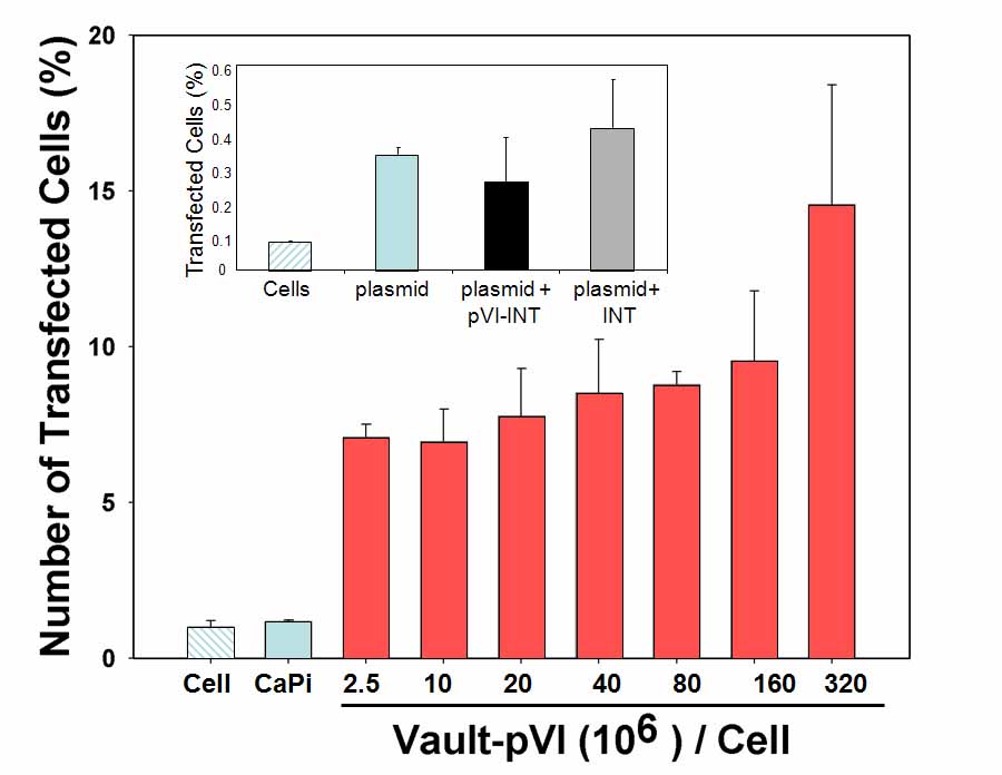
These findings indicate that vault particles can be modified to enhance the transfer of selected biomolecules across the cell membrane and they offer new avenues for adapting vaults for cell delivery of therapeutic molecules.