Big Kids: Delivery of Vaults to Cells
The green fluorescent protein been attached directly to the major vault protein, MVP. Even though this fusion protein (called GL-MVP) quite a bit larger than MVP, it still assembles into vault particles with surprising structural integrity. The GL-MVP vaults are highly fluorescent and thus can be visualized by fluorescence microscopy. Purified GL-MVP vaults have been added to cultured cells and their uptake by the cells with a special fluorescence microscope called a confocal microscope. An cell line (called HeLa cells) was used. Following a one hour incubation at 37oC the media containing the fluorescent vaults was removed followed by rinsing the cells and monitoring for fluorescence. Using this approach, it was shown that HeLa cells can readily take up vaults from the culture media. An example of HeLa cells that engulfed GL-MVP vaults is shown in the Figure.
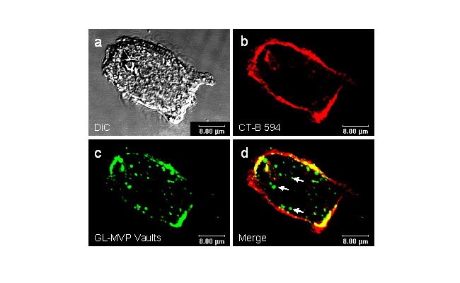
Figure (below). HeLa cells were incubated with GL-MVP vaults for 1h at 37oC, washed and incubated with a dye called CT-B 594 (this dye stains cell membranes). The cells were then washed and mounted on slides and visualized by confocal microscopy. (a) A regular light image showing the HeLa cell; (b) Mostly membrane staining (red) seen with CT-B; (c) Green fluorescent vaults seen on the membrane and inside the cells; (d) a merge of b and c showing vaults inside the cells (arrows). Fluorescent microscopy was performed at the UCLA/CNSI Advanced Light Microscopy/Spectroscopy Shared Facility. Figure is from Kickhoefer et al. (2005).