Kids: History

In the mid 1980s the Rome Laboratory at the David Geffen School of Medicine at UCLA was studying the movement of proteins from their site of production (an organelle called endoplasmic reticulum) to their destination (the lysosome). The proteins were shuttled in cells in a specialized container called a coated vesicle. Nancy Kedersha was purifying coated vesicles and examining the vesicles using an electron microscope (EM). One population of coated vesicles was contaminated with an unusual ovoid particle that was highly regular in its form and dimensions.
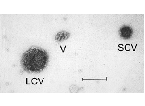
This figure shows an EM image from a coated vesicle sample. Present is a large coated vesicle (LCV), a small coated vesicle (SCV) and an unusual particle with a complex knotted structure (V). The bar represents 100 nmeters.
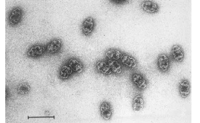
Dr. Kedersha modified the purification procedure and was able to separate the ovoid particles away from the coated vesicles. The purified particles are shown here. The bar (lower left) is 100 nmeters.
The purified particles were examined using biochemical methods which revealed that the particles were unrelated to coated vesicles and thus novel. Vaults was chosen as the name for the particle, a term selected to describe the shape of the particle which consists of multiple arches like those that form vaulted ceilings of churches.
